Trials and Tribulations in Medical Prepping

Frustated with preparing for end-of-life-as-we-know-it medical concerns?  Today a reader and health care provider shares her experience with trying to get her doctor to help her with medical prepping.   

* * *

I personally have been ethical and forthright with my personal health practitioners about preparing for medical disasters in a potential TEOTWAWKI scenario/s. I am sharing some personal experiences encountered here, which all relate to trials and tribulations of finding an MD who is a fellow prepper.

Here are three occasions with three physicians.

The sad reality is that the first, my primary caregiver, was a jewel in the rough, but she closed her practice in my locale and moved far away. Although she herself had not evolved yet to the actual practice of preparing herself or her family, she acknowledged the potential increase of my concerns to be personally prepared, and validated my requests for issuing me Rx’s for broad spectrum antibiotics for my medical storage preps. There was a long standing doctor-patient relationship here with us and one of mutual trust, professional and moral ethics. Of course, my medical competency and antibiotic use prudence was a key component in her granting this Rx transaction.

The next prescription request I made was presented to a specialist I was referred to. Our relationship was short, but none the less, I was prescribed some rarer, older meds due to a personal history of chemical allergies from most of the newer diuretic preparations. Thus, I had to ask this time for an extra Rx to fill for my storage preps of this newly added med. He flatly declined – even though he knows both my husband and I are medical practitioners! There was no further discussion from him on this issue. I thus had to find another specialist who would serve my needs to take care of my medication preps.

This brought me to the latest conundrum. I was referred to another primary physician, since mine had moved away. This time I knew to ask outright about his beliefs regarding Armageddon medicine and prepping before I even let him examine me on the initial visit. I was relieved to find an open mind and also a mutual mindset when it came to the practice of medical prepping. All went well until I became acutely ill with a near-syncopal episode in a public place (I knew I was having a severe allergic medication reaction and took measures to self-medicate with an antihistamine immediately), and was transported to the ER in tachycardia. 

Upon negative labs, ekg’s, and 8 hrs of waiting for the second set of neg CKMB’s and for my primary to end his office hours to come to see me, I was finally greeted by an unknown “partner MD” covering for my MD. My MD was off for an extended weekend. I had never met this MD before. He admitted me to the hospital. I objected, based on my personal knowledge of my medical history and that I had experienced this before, and treated myself with Benadryl and some O2, monitored my b/p and rested. We thus would also have to change medications again. He became outraged at my attempts to treat myself (which by the way worked), and gave me an ultimatum that if I had not agreed to the in-patient stay, that he would remove himself from my case and that I would have to sign out AMA.

You know, I have treated patients for 35 plus years, and I have never treated them with audacity. I even tried to reason with him, that I was going home with another MD, my husband, and we would be fine, and if I had any other untoward symtoms, my husband would have me back there in a flash. But, no. I have to stay. I felt entrapped. I was entrapped. I ended up asking again for the hospital administrator, who was also out of town, and instead was referred to the Sr. Nursing administrator, who was next in charge. She repeated the same words. They took the collaborative path of cohorts instead of listening to my plea. The negotiable offer was made to stay for a 24 hour observation. I complied and stayed and did not sleep a wink the entire night. I then had to wait until the 25th hour and still he never did come back to examine me. I called the administrator again to ask when I could sign myself out. He ended up writing a discharge over the phone, (verbal order), and I went home….quite angry, and had to start all over to find a new primary physician that would be compatible with my needs as a prepper.

One week later, I received a certified CYA letter from this MD, stating he was dropping me as a patient on the basis of Mistrust in Him. Great observational skills on his part!

What did I learn from this experience in reference to medical prepping? It is imperative to seek out by the reference of a friend or another MD whom you trust, to discuss if that physician or practitioner is a prepper themself. Keep searching until you find one that you can trust. Ask all the questions of whom will have access to your personal information and assigned to your care, in his/her absence. Can you sign a contract with your physician to be able to dispense a prescription to you, so you can self medicate when you clearly (and in my case, by license), know what you’re doing, and also agree to relieve that MD from medical harm, malpractice? The working relationship must be based on mutual understanding, respect and trust.

It will be difficult to find a fellow prepping MD, but not impossible. When you do find that MD, honor him or her by referring others to them.

– Anonymous 

Doc Cindy responds:  Anonymous has experienced many of the obstacles to medical prepping:  issues of trust, physician personality, inflexibility of the current system, and hidden concerns.  Why would a doctor be so unwilling to entrust a patient with her own care?  My suspicion is either this doctor has ego problems or he’s been burned before, i.e. – sued by a patient in a similar situation.  Health care providers functioning within “the system” must support the system or face losing their jobs.  Free-thinkers are more likely to be found in small or solo practice.

Related articles:  How to Get Your Doctor to Help You Stockpile Medicine


Enhanced by Zemanta
Posted in Medical archives, Perennial Favorites, Stockpiling medical supplies, Stockpiling medications | Tagged , , , , , | 4 Comments

Photo Quiz Question – Q.001

Photo Quiz Question – Q.001 – June 10, 2011

As this web site has developed I’ve continually tried to adapt it to the interests of my readers.

At medical conferences, photo quizzes are always popular, and at the-end-of-the-world-as-we-know-it, diagnosis will be largely clinical, that is, based on history and physical exam, rather than testing or X-rays.

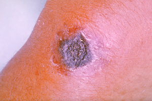
So what does the above image depict and how is it treated?

To be honest, submit your response in the box below before turning to the answer.

For the correct answer, CLICK HERE.

Posted in Medical archives, Perennial Favorites, Photo Quiz Questions, Skin | 5 Comments

Photo Quiz Answer – Q.001

Photo Quiz Answer – Q.001 – June 10, 2011

Cutaneous anthrax due to Bacillus anthracis.

Most cases of cutaneous anthrax are caused by contamination of an open wound, often with bacteria present on wool, leather, or hair from infected animals, particularly goats.

Untreated, 20% of patients with cutaneous anthrax will die, though death is rare with appropriate antibiotic therapy (<1%).

Ciprofloxacin 500 mg twice daily or doxycycline 100 mg twice daily is recommended for the treatment of anthrax in adults and children (with the consideration that amoxicillin or penicillin may be preferable in growing children or pregnant women.)

Treatment should be continued for 10 days, unless a bio-terrorist attack is a concern, in which case 60 days of treatment is recommended.

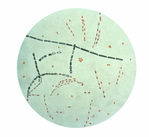
The above image is a Gram stain of the anthrax bacterium.  For additional images, see below.

For more information, visit Questions and Answers About Anthrax

Anthrax

Image via Wikipedia

Photomicrograph of Bacillus anthracis from an ...

Image via Wikipedia

Gram-positive Bacillus anthracis bacteria (pur...

Image via Wikipedia

Enhanced by Zemanta
Posted in Anthrax, Anthrax, Education, Medical archives, Photo Quiz Answers | Tagged , , , , , , , | 3 Comments

Week 14 – Question of the Week: How do you wish your doctor would help you prep?

Week 14: 2011-06-9

Today I’m asking all readers to comment on this question:

How do you wish your doctor would help you prep?  Are there any concerns beyond stockpiling medicine?

Have you tried talking with your doctor?  Are you afraid to even broach the subject?  Has anyone tried my advice on getting your doctor to help you stockpile medicine?

Check back soon and see what your fellow readers have to say.

Image – gonococcal eye infection of the newborn

7 Comments

Who makes the rules?

A common theme in the comments I receive is frustration over preparing for future medical events when one lacks the license to do so.

Why makes the rules, anyway?

Is is fair that only doctors can prescribe antibiotics?  Does the government really care about our welfare?  Or are health laws an impediment to responsible citizenship?

Six months ago my son broke his femur and underwent surgery.  I’m pretty sure I’m glad his doctor was not only experienced, but licensed.  It adds a level of confidence.

But then, it seems ridiculous to me that hearing aids cost thousands of dollars, when the iPad my daughter wants is under $600 (with the lime-green case).

Or why should I pay $200 for eyeglasses when you can get cheater-readers for a few bucks at the Dollar Store?  Who made those rules?

Take antibiotics, for example.  Should a person need a license to prescribe amoxicillin? As one reader said, how hard can it be to take amoxicillin for a sinus infection? That would be a healthy person talking, one who hasn’t experienced C. diff, or MRSA, or penicillin-resistant pneumonia, or meningococcal meningitis, someone who hasn’t watched a baby die of Haemophilus influenzae or pertussis. Most people are healthy, and only take antibiotics for minor respiratory, urinary, or skin infections.  They haven’t really considered the life-saving role antibiotics can play, nor the life-endangering side-effects they can cause, nor the issue of antibiotic resistance due to wide-spread use.

The focus here is not antibiotics, but rather the question of regulation of health care. Regulations can help, regulations can hurt.  The frustration for preppers is primarily that of lack of access to medicine that may be life-saving in the future.  If no doctor is available, isn’t it only fair to be able to treat yourself with something that might help rather than follow the rules and possibly die? I side with the first option.

Education is the key to taking care of oneself and one’s family.  To that end I’m doing what I can to assist.  My book’s nearly done – the first edition anyway.  How does one cram everything a person might need to know into a single volume? It can’t be done, but I’ve got to stop somewhere (and my kids have to eat, too).  So far I’ve dedicated a thousand hours to the effort.

I’ve always been one to follow the rules, at least most of them (no speeding tickets, though I’m not above jaywalking), but the truth is, you don’t need a medical license to do much of what doctors do.

Is there some way I can assist, some question you may have? I’ll try to give an answer for times when the rules won’t apply.  In the meantime, I’ll respect regulations that are primarily intended for our own good.

Enhanced by Zemanta
Posted in Medications, Perennial Favorites, Preparation, Prescription Medications, Public health | 2 Comments

Week 13 – Question of the Week: How far along are you in your medical prepping?

Week 13: 2011-06-2

With 2012 approaching, today I’m asking all readers to comment on this question:

How far along are you in your medical prepping?

From comments I’ve received, most are just now beginning.  A few, less than 5% by my estimate, have supplies for as much as a year.

What are the costs?  What are the challenges?  How much time is involved?

Check back soon and see what your fellow readers have to say.


Image attribution: By Cienkamila (talk · contribs); slightly edited by Odder (talk · contribs) [CC-BY-SA-3.0 (www.creativecommons.org/licenses/by-sa/3.0) or GFDL (www.gnu.org/copyleft/fdl.html)], via Wikimedia Commons

Enhanced by Zemanta
Posted in Medical archives, Perennial Favorites, Preparation | 5 Comments

Where to begin?

Where to begin? As this blog develops, specific advice on relevant topics will be added on a regular basis.  If you have comments or questions on a particular aspect of health care, please enter them below.             

In the meantime, start now by educating yourself and stockpiling your medical kit with inexpensive over-the-counter medications and equipment. 
  
How will you know what to get and how to effectively use your supplies? 
  
One place to start is with my recently released book, 101 Ways to Save Money on Health Care.  You don’t even have to buy it – just check it out from your local library. 
  
Although the book was written with economics in mind, since knowing how to care for your own health is an important aspect of both Armageddon Medicine and saving money on health care, this is an inexpensive and timely starting place.  It explains how to treat over 30 common medical conditions, often without consulting a professional.    Continue reading
Posted in 101 Ways / Health Care, 101 Ways to Save Money on Health Care, Ankle sprain, Arthritis, Back sprain, Blood pressure meds, Books, Bookshelf, Chronic diseases, Diabetes Mellitus Type II, Diseases, Immunization, Injuries, Lacerations, Medications, OTC Pain meds, Over-the-counter meds, Perennial Favorites, Preparation, Prescription Medications, Save Money, Slide show, Sprains, Stockpiling medical supplies, Stockpiling medications, Vaccination | Tagged , , | 4 Comments

Clinical Laboratory Procedures under Austere Conditions: Part II – Light Microscopy

The following post on clinical laboratory procedures is contributed by Pete Farmer,  who holds advanced degrees in research biology and history, and is also an RN and EMT.  For Part I of the series, click HERE

* * *

As outlined in the first installment of this series, adopting an expeditionary or operational mindset can be enormously valuable for anyone interested in medical care under austere conditions. We noted that special operations medics, humanitarian aid workers, and others who practice healthcare in extreme environments have much to teach us about delivering care under less-than-ideal conditions. As previously noted, first-world medicine has evolved and depends upon an intricate high-tech infrastructure designed to provide services and support for the physician or other first-line provider. Among the most important of these services are clinical laboratory tests. In the field, however, you may have to depend upon only those resources available in your immediate surroundings. In all likelihood, that means having fewer laboratory tests and diagnostic procedures available as adjuncts to clinical decision-making. This is the bad news. The good news is that, using some fairly simple tools, one can perform some extremely useful laboratory tests and procedures, even under hardship conditions.

Among the most important items to be found in a field clinical laboratory is the basic light microscope. This humble and commonplace device, essentially unchanged from the time of Robert Koch and Louis Pasteur, and familiar to most anyone who has taken a high school or college introductory biology course, is a powerful clinical tool when employed properly. Rather than reinvent the microscope piece-by-piece for the following discussion, the reader is assumed to have a basic knowledge of what a light microscope is and how it functions (Note: readers in need of an introduction to or a refresher in basic light microscopy are directed to the links and notes at the end of this article).

For the purposes of the following discussion, we will use a light microscope similar to that in Figure 1, as our prototype. This design was state of the art in the 1930s, and is still used widely by students and other microscopists – although newer examples benefit from improvements in materials and workmanship. Please note that this design – a Zeiss – does not have a fixed electric light source, but uses a mirror to gather and reflect ambient light onto the specimen. Choose the type best-suited to your needs – powered or unpowered. If you will be using the microscope where you will not have access to a reliable source of electricity, the latter may be indicated.

Most light microscopes are compound, i.e., employ multiple ground-glass lenses to refract (bend) light to achieve magnification (enlargement) of an image. The magnifying capability of a compound microscope is the product of the magnification of its individual ocular and objective lenses. The former, also called the eyepiece, is the lens nearest the eye and typically has 10x (tenfold) magnification. The latter, called objectives, are the lenses nearest the specimen, and magnify at various selectable ranges, generally 4x, 10x, 40x and 100x power, or some combination thereof. Bench-top light microscopes generally do not magnify at greater than 1500x, except for specialized, non-standard applications not relevant here. Total magnification is determined by multiplying the ocular lens power times the objective lens power, i.e., a 10x ocular lens paired with a 100x objective gives a thousand-fold magnification (1000x).

Another important principle of microscopy is resolution, defined as the degree to which detail in the magnified image is retained. It is often described as the ability to distinguish between two points. In layman’s terms, resolution is the degree of detail which is visible and sharply delineated. Having 1000x magnification is of little use if the resultant image is blurred and not crisp-enough to allow visualization of fine detail. The maximum useful resolution of a typical light microscope is typically around 200 nanometers (1nm = 1 nanometer = 1×10 -9 meters), or slightly less than the size of most bacterial cells. Therefore, properly-used, a standard light microscope can be used to visualize most bacteria, protozoans, algae and fungi. Viruses cannot be visualized using std. light microscopy. Resolution (R) is dependent upon the wavelength of light passing through the specimen (slide – specimen – cover glass), as well as the properties of the lenses used in the microscope. The smaller the resolution number (value) of a microscope, the finer the detail visible. Bench-top light microscopes employ light in the visible or near-visible spectrum; blue light (wavelength 400 nm) – which has a shorter wavelength than red light (700nm) – gives better resolution than red light. Blue light is obtained by inserting a blue filter over a visible light source or by using a lamp which emits blue light.

Resolution and focus are important not only laterally, between adjacent points in a specimen, but vertically – along the long axis of the microscope. This characteristic is most often referred to depth-of-field. This construct, familiar to anyone experienced in photography, refers to the distance between nearest horizontal plane in focus in the foreground and the furthest-away horizontal plane in focus in the background. Inside the depth of field, the image is resolvable and in focus; outside of the depth of field, it is not. Depth of field is dependent upon numerical aperture and magnification of the objective lens; i.e. a 4x objective with numerical aperture of 1.0, has greater depth of field than a 100x objective with 0.95 aperture. Most often, one is trading less depth of field in return for greater magnification.

Numerical aperture (NA) value determines the degree of useful magnification of a given lens. It is dependent upon the refractive index (n) of the medium between the specimen and the front of the lens, and the angle of the most oblique light rays entering the objective lens. Air has a refractive index of 1, which limits resolution. However, the value of NA (useful magnification) can be increased by putting immersion oil between the cover glass on top of the specimen and slide, and the objective. Immersion oil has a refractive index (~ 1.5) more closely approximating glass than air, which allows higher magnification and better resolution. Using a 100x objective with immersion oil gives a useful magnification of approximately 1,500x, sufficient to view most bacteria. Most protozoans, algae, and fungi may be visualized without oil immersion. 

Contrast is needed to distinguish an object from its background. Since microbes themselves are composed of a high percentage of water, and are usually viewed in aqueous (water) solution, a means of increasing the contrast between the specimen and its background is usually employed during specimen preparation. Typically, a chemical stain or other colored reagent is used for this purpose. Many staining techniques are more than a century old, but remain among the most commonly-used and effective means of visualizing and identifying microbes, tissue samples and other specimens.

A simple staining procedure employs a single stain, and generally imparts the same color to all cells and structures, regardless of type. The stain increases contrast by either coloring the cells but not the background, or the reverse – staining the background but not the cells themselves. The charge (positive + or negative -) of the stain molecule (chromophore) itself is also important. Most biological specimens are charged either positively or negatively (or both, in different regions). Similar charges repel, dissimilar charges attract. The stain methylene blue is positively-charged and basic (has a pH greater than 7), and thus adheres to the negatively-charged outer surface of certain types of bacteria. Nigrosine is an example of an acidic (pH less than 7), negatively-charged stain; it is repelled by negatively-charged bacteria and stains the background instead.

Differential staining employs a multi-stage staining process which utilizes the unique staining characteristics of different microbial cells and/or structures. Typically, a stain is used, and then “fixed” (made permanent) by use of heat or chemical treatment – and then washed off using the appropriate solvent. Counter-staining (staining with a different colored reagent) allows the microscopist to provide further contrast and differentiation with the background and the objects stained in the first-pass stain. The Gram Stain (to be covered further on in this series) is the most commonly used differential staining technique in microbiology.

Specimen preparation is exceedingly important for successfully microscopy; without it, the best equipment will not function optimally; with it, even modest equipment can perform well. Recall that successful visualization and magnification of the image being visualized depends on either reflected or transmitted light. The former uses a light source placed at an angle, and illuminates the specimen, which is then visualized. Anyone who has looked at a three-dimensional object under a stereoscopic microscope is familiar with this sort of light source, which is not all that different from indirect lighting used by a studio photographer. Transmitted light, on the other hand, must pass through the slide, specimen and cover glass (or slip) to reach the objective lens of the microscope. A light source directly below the stage (see Figure 1) is used, either an electric light or a mirror. Given the considerations of depth of field, thickness and uniformity are important considerations for any specimen which must be cut into extremely thin sections for viewing. A precision instrument called a microtome is used for this purpose. It is important to note that in the field, tissues requiring extensive preparation, such as multi-stage thin-sectioning or staining, are generally not examined because of the extensive equipment and supplies needed. Luckily, many useful microscopic preparations do not require such sophisticated methods. Many clinically-useful microscopic examinations of blood, sputum, saliva, urine and stool can be carried out in the field using simple sample preparation procedures.

Next installment, we’ll continue our examination of light microscopy with a discussion of sample preparation and some basic staining techniques.  

Figure 1: Zeiss Laboratory Microscope (c. 1879)

File:Microscope Zeiss 1879.jpg

(For additional images see: http://www.olympusmicro.com/primer/anatomy/introduction.html)

  1. http://www.ekcsk12.org/faculty/jbuckley/lelab/microscopeuselab.htm

This article is basic, but explains a typical student model microscope well

  1. http://science.howstuffworks.com/light-microscope.htm

This is a well-done survey which takes the reader from the basics through more advanced designs. You may by-pass the sections on fluorescence microscopy and other specialized subtypes. 

  1. http://www.olympusmicro.com/primer/
  2. http://biology.fullerton.edu/facilities/em/IntroLightMicro.html

Explains the physics behind light microscopy, including a well-done section on optics.

  1. Carl Zeiss Microscopes – Zeiss is one of the most respected optical instruments firms in the world; Zeiss.com has a selection of bench-top light microscopes. There are many other reputable firms as well.
  2. http://www.microscopyu.com/articles/formulas/formulasfielddepth.html

Discussion of depth of field in microscopy, with calculations.

 Note: Readers unfamiliar with or in need of a refresher in the basics of light microscopy may benefit from the following: (a) an introductory course in biology, with laboratory or (b) an introductory course in microbiology or bacteriology, with lab (c) some scientific supply houses sell student microscopy kits designed for at-home use; these include basic instructional materials and introductory projects; (d) there are also many excellent texts and websites available.

Reference cited: Atlas, Ronald M., Ph.D. “Principles of Microbiology,” Mosby: St. Louis, 1995.

Copyright © 2011 Peter Farmer

Posted in Contributors, Equipment, Field equipment, Medical testing, Microscopic examination, Perennial Favorites, Pete Farmer | Tagged , , , , , , | Leave a comment

Memorial Day Through the Eyes of a Veterinarian and a Veteran

Today’s post is contributed by one of our professionals, both a veterinarian and U.S. veteran.

* * *

Flag of the United States Navy

Image via Wikipedia

I have been blessed to have had a full life with two great daughters and many friends on this and other continents; to have been able to serve the health of animals and their caretakers in the communities in which I have practiced; to have been an active member of law enforcement, churches, fraternal orders and service organizations in a number of parts of this great country. I have done so because I have had the life and FREEDOM to participate. That freedom was bought by the sacrifices of other members of the armed forces, many of whom did not have the opportunities I have had since they didn’t return from service or were returned in flag-draped coffins.

We are “the land of the free and the home of the brave” and it is my hope that as Memorial Day approaches, you will pause for a moment and reflect on those freedoms that we possess that are not available anywhere else on the globe. There are those who are viciously jealous of the United States and who would take those freedoms from us if we let them have the opportunity. As I write this, there are men and women serving “in country” and in their homeland to protect those freedoms from the forces of evil that would violently strip them from us if given the slightest chance.

My Dad served as a skipper of a number of Landing Craft in the Pacific during WWII. He survived the Battle of Midway, Wake Island, the Marianas and others to come back to a teaching career in the inner city schools of northwest Indiana. Few understood why he was such a quietly strong man and why he put up with the educational system as it was…and to which he contributed so much. I think if you will look around at those who have raised their right hands and took the oath “…to defend the Constitution of the United States against all enemies, foreign and domestic…” you will find other men and women quietly serving their communities who have experienced wars and conflicts while wearing the uniforms of the United States armed services. For these silent warriors and to those who have made the ultimate sacrifice for our great country it is my hope that you will remember them on Memorial Day; that you will say a thankful prayer and one for protection of those currently serving.

It is also my hope that you will feel emotion when the Stars and Stripes pass in review, with an understanding what it truly means to be an American.  So, if you’ve served, salute Old Glory as she passes in review. If not, place your hand over your heart in reverence of the freedoms she represents and in recognition of those who served so that you could have those freedoms.  God bless America!

Image via Wikipedia

Always Faithful, Doberman, Military Working Dog, MWD, World War II Memorial, War Dog Cemetery located on Navel Base Guam

(Above flag image was released by the United States Navy to the public domain with the ID 030626-N-1539M-002.)

Enhanced by Zemanta
Posted in Disaster Relief, Perennial Favorites, Public health, Veterinary, Volunteering | Tagged , , , , | Leave a comment

Week 12 – Question of the Week: How can we best serve our country in preparing for an Armageddon event?

Week 12: 2011-05-26

With Memorial Day approaching, today I’m asking all readers to consider this question:

How can we best serve our country in preparing for an Armageddon event?

I know several readers and contributors are serving or have served in the military.  Many are concerned about the direction our country is going.  Some feel the government actually interferes with individual preparedness. 

What are your feelings, concerns, and thoughts on the question?

Check back soon and see what your fellow readers have to say.

– Doc Cindy
Enhanced by Zemanta
Posted in Medical archives, Perennial Favorites, Preparation, Public health, Question of the Week | Tagged , | 9 Comments