Thyroid disease – Hypothyroidism (low thyroid) – Part 1 of 5

The following is excerpted from my upcoming book, Armageddon Medicine.

Thyroid disease – Hypothyroidism (Low thyroid)

The question of treating low thyroid in a post-apocalyptic world is a common one.  If Synthroid becomes unavailable, is a hypothyroid person doomed?

The short answer is:  likely not – so read on.

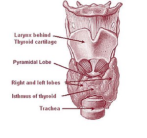
Overview of the thyroid system (See Wikipedia:...

Image via Wikipedia

Hypothyroidism (low thyroid, or thyroid insufficiency) encompasses a wide spectrum of problems, from asymptomatic (subclinical) hypothyroidism, to severe disease and coma.  Only once in my 30-year medical career have I seen a patient obtunded from hypothyroidism, and I’ve never seen a patient with an iodine-deficiency goiter, though that disease may resurface after global catastrophe.

For simplicity sake, I’ll divide low-thyroid disease into two main classes: those with an absolute deficiency of thyroid hormones and those with a partial deficiency.  People with no thyroid hormone whatsoever must receive replacement therapy or they will gradually succumb to severe thyroid deficiency (fatigue, low heart rate, slowed thinking, swollen legs, dry skin, hair loss, and possible coma and death).  People with a partial deficiency may live for years or even decades with minimal or no symptoms.

The real question is: which sort of patient are you?

Most people won’t know the answer to this question.  You can, however, determine the answer with a little investigation.

Absolute thyroid deficiency is mainly caused by two conditions:  severe, intrinsic thyroid disease or as a result of treatment for hyperactive thyroid (high thyroid, Grave’s disease).  Patients who have undergone total thyroidectomy or total thyroid ablation via radiation for a hyperactive thyroid should, theoretically, have no remaining functional thyroid tissue.  This is not always the case, since some doctors treat hyperthyroid disease with a sub-total thyroidectomy or incomplete irradiation, aiming to leave sufficient thyroid tissue to avoid the need for replacement therapy.

Much more common is partial thyroid deficiency.  To determine this, what doctors actually measure is the serum TSH level (thyroid stimulating hormone).  TSH is produced by the pituitary, and as the name implies, TSH stimulates the thyroid to make thyroid hormone via a fine-tuned feedback system.  If your serum thyroid level drops a bit low, the pituitary responds by producing more thyroid stimulating hormone, which in turn causes the thyroid gland to increase output of thyroid hormone (assuming there is thyroid tissue to stimulate).  If your serum thyroid hormone level increases, TSH levels drop.

Nowadays most patients with hypothyroidism are diagnosed by an elevated TSH level rather than by symptoms.  Sure, a patient may complain of fatigue, but who isn’t tired?  I’ve had many patients with a mildly elevated TSH level who were placed on thyroid replacement therapy, either by myself or another physician, who felt absolutely no different taking Synthroid.  A serum TSH level is a common screening blood test for middle-aged and older people undergoing a routine exam.  Therefore mild hypothyroidism is thus often identified in patients who had no clue anything was wrong.  Another name for this problem is ‘subclinical hypothyroidism,’ that is, too mild to cause symptoms.

The normal range for TSH varies from lab to lab, but is approximately 0.3 to 5.0 mIU/L.  Patients are commonly placed on thyroid replacement therapy when their TSH is elevated a few points, even if they have no symptoms.  However, this practice is controversial.  Per a review article in The American Family Physician, “In current randomized controlled trials, levothyroxine replacement therapy for subclinical hypothyroidism did not result in improved survival or decreased cardiovascular morbidity. Data on health-related quality of life and symptoms did not demonstrate significant differences among intervention groups. Some evidence indicates that levothyroxine replacement improves some parameters of lipid profiles and left ventricular function.” (Am Fam Physician. 2008 Apr 1;77(7):953-955, online at: http://www.aafp.org/afp/2008/0401/p953.html)

My own experience confirms this data.  So if you won’t feel better or live longer, and if you are not experiencing symptoms of hypothyroidism, patients in this category probably don’t require treatment.

Thyroid

Image via Wikipedia

However, there are patients whose TSH levels are elevated by 20, 30, or even 100 points.  These patients are likely to exhibit symptoms and on blood testing are likely to show decreased levels of serum thyroid hormone.  Common symptoms of hypothyroidism include:  weakness, dry or coarse skin or hair, fatigue or lethargy, swollen eyelids or face, feeling cold, slowed speech or thinking, and constipation.  Of course, many patients have one or more of these symptoms and do not have any thyroid disease.  However, patients with decreased thyroid function will improve if given thyroid supplementation.

Future installments of this topic will include:

  • Old, current, and future treatment of hypothyroidism
  • Figuring out what type of thyroid disease you have
  • Stockpiling medication
  • Alternative and complementary medicine

For more answers to your thyroid questions, see HYPOTHYROIDISM – Answers for the End of the World.

Enhanced by Zemanta
Posted in Blood tests, Fatigue, Hyperthyroidism, Hypothyroidism, Medical archives, Medical testing, Perennial Favorites, T3 and T4, Thyroid disease, Thyroid tests, TSH, Weight gain, Weight loss | Tagged , , , , , , , | 7 Comments

Photo Quiz Question – Q.003

Photo Quiz Question – Q.003 – July 1, 2011

31-year-old white male, circa 1951, missionary to Nigeria

Illness started with fever and aching for 2-3 days, followed by development of blistering rash.  Above image depicts rash a week into the illness.

What does the above image depict?  Is there any treatment?  Should the patient be isolated?
 

To be honest, submit your response in the box below before turning to the answer.

 Photo courtesy of Wikimedia Commons

Enhanced by Zemanta
Posted in Acute diseases, Education, Medical archives, Perennial Favorites, Photo Quiz Questions | Tagged | 13 Comments

Week 16 – Question of the Week: Bioterrorism – Worth worrying about or not?

Week 16: 2011-06-30

I’m completing the chapter in my book on bioterrorism and would like to ask everyone: how concerned are you?

Have you done anything to prepare in the event of a bioterrorist attack?  Do you worry about receiving a letter with a suspicious powdery substance? 

Is bioterrorism really worth worrying about on an individual level?

Comments, questions, and suggestions are all appreciated.  Submit your response in the block below.

 – Doc Cindy

 Image:  Anthrax targets

Graphical depiction of 2001 anthrax mailing (s...

Image via Wikipedia


Enhanced by Zemanta
Posted in Anthrax, Bioterrorism, Education, Medical archives, Perennial Favorites, Preparation, Public health, Question of the Week | Tagged , , , , | 3 Comments

Home birth – One doctor’s experience

It didn’t happen on purpose.  Not exactly, anyway.

This was child #3, during my stint in Appalachia with the National Health Service Corps.  Though I lived in the biggest town around (with a grand 5,000 population), no doctor was willing to allow a VBAC birth (vaginal birth after Caesarean).  Mind you, I’d already had one without incident.  Child #1 was C-section for failure to progress, but Child #2 was a normal labor and delivery.  So what was the big deal?  I never received an answer.

Instead I found an OB/gyn in a larger town an hour and a half away, who was willing to accommodate my request.  I made regular monthly, then weekly visits, throughout my normal pregnancy.  I figured I’d have plenty of time to reach the hospital.  I’d been in labor nearly 24 hours with each of the first two babies.

Turns out, I was wrong – fortunately.  I called my husband, who was out-of-state, at 6 a.m., asking him to hurry home.  He was less than six hours away (five, at 80 mph), and should be back in plenty of time.

It was a warm day in May, the kids were out of school, so my daughter was able to watch her younger brother (who was cooperating for some reason).  When the contractions grew stronger, I took refuge in our over-size bathtub, which was large enough to float in.  This was far more relaxing than a hospital bed, surrounded by strangers.

Was it the environment or the baby?  I’ll never know. But labor progressed more quickly than expected.  After about 6 hours I started growing irritable – angry, really.  Why wasn’t he home yet?  I’m not sure I recognized this as transition labor, but I knew I wasn’t going to any hospital.

Suddenly, he burst through the door.  “I’m calling an ambulance,” he cried, heading for the phone.

“I’m not going anywhere,” I retorted.  “Get over here and help me.”

Between contractions I managed to make it to my bed.  Ten minutes later, my precious daughter was born.  Though he would never have chosen to, my husband was delighted to help deliver the baby.  He even videotaped the placenta.

My friend, Dr. Steve, stopped by a few hours later and placed a few stitches (though I’m not sure I really needed them).  By then everything was cleaned up and the baby was happily nursing.  It felt odd to resume life so immediately, without the hullabaloo of the hospital.

It also seemed strange that when I filed for a birth certificate, the health department said they hadn’t heard of a home birth in years – and this deep in Appalachia.  I still find it hard to believe.

That experience convinced me that a home birth was far preferable to a hospital delivery, at least for a healthy mother and child.  I would have repeated the experience with my fourth, but things didn’t go as smoothly.  His big head got stuck and I’m not sure he would ever have delivered without another C-section.

In retrospect, 31 years after the first delivery, I think child #1 would have delivered, had we waited a little longer, and maybe had circumstances been more relaxed.  Child #2 and #3 clearly were home-deliverable.  However, without intervention, I wonder if trying to deliver child #4 might have killed us both.

The topics of prenatal care, labor, and delivery have not yet been addressed in Armageddon Medicine.  I need more hours in the day to get everything done.  I know we have some obstetricians and midwives on board, and hope they’ll be willing to help out, maybe write an article or two . . . perhaps while waiting for the next child to be born.

Copyright © 2011 Cynthia J. Koelker, MD

Enhanced by Zemanta
Posted in Complications, Delivery, Home birth, Labor, Medical archives, Newborn care, Perennial Favorites, Pregnancy | Tagged , , , , , , , | 1 Comment

Week 15 – Question of the Week: How are you encouraging your neighbors to prep?

Week 15: 2011-06-23

This week I was talking with Pastor Bill, who once told me he’d stocked over 300 rolls of toilet paper.  Today I asked him what other preparations he’d made, which turned out to be quite a few – a generator, an outhouse, wood stove heating, a well, food supplies.

Then he went on to add that he thought he was the only one on his road making any preparations at all.  What about the newlyweds across the street who are expecting a baby soon.  What are you going to do, turn them away?

This got me wondering what you advanced preppers are doing to encourage your neighbors, friends, and relatives to prepare.  We can’t all bug out.  I might be able to garner enough supplies for my extended family, but I cannot supply the entire community.

So what are you doing to encourage your neighbors, friends, and relatives to prepare?  And what success have you had?

Anyone is welcome to submit an answer to share.

Enhanced by Zemanta
Posted in Medical archives, Perennial Favorites | Tagged , , , , , | 6 Comments

Survival Gardening with Doc Cindy

When my vision blurs from typing, I head for my survival garden.  I hope I survive the season.

Yellow onions

Image via Wikipedia

Writing a book on Armageddon Medicine will change your view of the world.  For instance, when I see a patch of grass, I’m overcome with the urge to plant more onions.  In Ohio, you can’t plant tomatoes in April.  I started early, and since they will survive a spring snow, I planted onions – in my flower beds, in my landscaping, in my garden – more onions than I’ll ever eat.  The kids don’t like onions.  The dogs don’t like onions.  It’s up to me to consume them all, or perhaps barter for apples or wheat.

Farming is a lot of work – if I have to survive on what I can raise, I’m in trouble.  Doctoring is definitely easier – at least less physically demanding.

I live in inner suburbia, and counting the usable land at my house and my office, I have perhaps half an acre.  So far I haven’t planted onions in the front yard at my office – but I could.  What a waste of energy to keep mowing the grass when I could be harvesting wheat.

Unusual strains of maize are collected to incr...

Image via Wikipedia

Corn grows well in Ohio.  It’s growing all over my yard.  I’m not sure if it’s from critters who buried it in the winter or from my son throwing corn cobs in the lawn, which I don’t understand either (except that he’s a boy).  The volunteer corn inspired me to plant rows of corn.  I hate walking in the mud so planted rows in my lawn.  My neighbor fears I’ve gone off the deep end. Eating corn is so much more satisfying than eating an onion (but what will I do with all those stalks???)

I call myself a YouTube gardener – and have learned all about planting corn and potatoes.  Yummy carbs – “bad” now, but good for Armageddon.  My children will starve if they have to live on tomatoes, peppers, and cukes.   So now we’ve been mulching potatoes, no doubt spending more on straw than the potatoes will be worth.

Then there’s the stevia, an interesting plant that so far is more of a conversation piece than a useful sweetener.  Can we survive without sugar?  Doubtful.  Maybe it’s not too late for sugar beets.  Or maybe I should learn more about tapping a sugar maple.  It’s all so much WORK!  Can’t I just plant some Milk Duds and harvest them in a month or two?

In addition to vegetables, I’m reaping arthritis, tendonitis, and neuritis.  No wonder women died at 50 a century ago.  Their parts were all worn out.

I can’t be saving any money on this project.  My kids tell me to just go buy groceries.  I tell them the story of the Little Red Hen.  If they won’t help me plant the onions, or harvest the onions, or wash the onions, or cook the onions, then I’m not sure I’ll share my onion rings.

Tomato slices.

Image via Wikipedia

Enhanced by Zemanta
Posted in Medical archives, Perennial Favorites | Tagged , , , , , , , | 9 Comments

Photo Quiz Answer – Q.002

Photo Quiz Answer – Q.002 – June 17, 2011

Here’s a little more (fabricated) history to help you decide.

This picture was taken 36 hours after a Labor Day picnic, which this girl enjoyed thoroughly, lying in the grass, talking on her cell phone.

If you’ve ever suffered the problem, you’ll recognize it now – chiggers – the super-itchy rash produced by exposure to the harvest mite.

By the time the rash appears, the tiny mite is (usually) long-gone.  The rash occurs wherever a chigger has injected enzymes to dissolve the skin (all the better to eat you, my dear).

Treatment is aimed at prevention and treating the itch with Calamine lotion, topical steroids, and/or oral antihistamines such as Benadryl, Zyrtec, Claritin, or generic equivalents.

Eventually the spots go away even with no treatment, though it may take 2-3 weeks for the lesions to resolve.

Scratching may lead to a secondary bacterial infection, that might require topical or oral antibiotics.

No quarantine is needed, though it may be a good idea to wash clothing that may have been exposed to mites living in long grasses during the late summer months.

For more information, visit: emedicinehealth

Posted in Bites-insect-bug-tick, Education, Injuries, Medical archives, Photo Quiz Answers, Rash, Symptoms | Leave a comment

Photo Quiz Question – Q.002

Photo Quiz Question – Q.002 – June 17, 2011

It’s summertime . . . better watch out for this problem. Anyone can get it – no one is truly immune.

What does the above image depict?  Is there any treatment?  Should the patient be quarantined?

To be honest, submit your response in the box below before turning to the answer.

For the correct answer, CLICK HERE.

Photo courtesy of Wikimedia Commons

Posted in Education, Insect-bug-tick bites, Medical archives, Perennial Favorites, Photo Quiz Questions, Rash | 11 Comments

Week 14 – Question of the Week: Hyperinflation – is it coming to America?

Week 14: 2011-06-16

Hyperinflation – is it coming to America?

Today I’m asking your opinion on hyperinflation.

Should I buy a wheelbarrow to tote useless dollars?

How will health care be effected in the short-term?

Is Glenn Beck crazy or is hyperinflation around the corner?

Readers of this blog are clearly more concerned about the future of this country than those who haven’t even thought about prepping.

Please share your thoughts with me and our thousands of readers.

– Doc Cindy

Enhanced by Zemanta
Posted in Medical archives, Perennial Favorites | 4 Comments

Clinical Laboratory Procedures under Austere Conditions: Part III – Sample Preparation

The following post on clinical laboratory procedures is contributed by Pete Farmer,  who holds advanced degrees in research biology and history, and is also an RN and EMT.  For Part I of the series, click HERE.

Thank you, Pete, for this practical series.

* * *

Once you have obtained a light microscope and familiarized yourself with it, you are ready to do some simple sample preparation and staining techniques. Before beginning, remember that microscopy is a mature scientific and technical field with a sizeable body of knowledge that takes many years of dedicated and diligent study to master. Know your limits, and don’t hesitate to ask for assistance from a specialist or expert when you need it.

Sample preparation is essential to obtaining useable, rigorous, and reliable results. As stated in a previous installment of this series, the finest optical equipment is of little benefit if the sample is improperly prepared; conversely, modest equipment can out-perform its price if the specimen to be visualized is prepared carefully and with attention to detail.

The following discussion pertains (unless noted) to preparations involving slides, i.e., mounting, fixation, staining, and cover-slipping of a sectioned specimen on a glass slide. Thus prepared, the slide is placed upon the microscope stage, from which it can be visualized. Before beginning, the microscopist will need to assemble the necessary supplies. Typically, these are obtained from a scientific supply house, such as Carolina Biological Supply, Fisher Scientific, or similar. What supplies you require will depend on what microscopy procedures you wish to perform. Microscopy for general/basic science (see endnote) applications can be ordered from the above suppliers; materials for specialized clinical applications of the kind done by a pathologist may require a different supplier. Some of the many variables affecting your choice include the following:

What material are you preparing?

How thin does your section (thin slice) of material have to be in order to be magnified successfully? Remember, light must be transmitted through the section for visualization to occur (if you do not know the answer, you should consult a reference).

Does the material to be sectioned require any pretreatment in chemical solution or other advanced preparation? Some materials are too soft or too porous to section properly unless pretreated.

Does your procedure require a specimen of uniform thickness? If so, how do you plan to assure such thickness?

Does your specimen require immobilization upon the surface of the slide, so that it does not move during visualization? Remember, at extreme magnification, small movements of your specimen may displace it from the field of vision.

Does your specimen require chemical (use of a chemical agent which bonds with the constituents of a cell or tissue and “fixes” or holds them, in place) or heat (use of heat for the same purpose) fixation?

How many sections (slices) of the specimen are required? Must they be of uniform thickness and dimensions? If so, you may wish to invest in a microtome, a device for cutting extremely thin slices, or sections, for use in microscopy. For simpler tissue preparations, a razor is often sufficient. For some types of samples, such as aqueous (water) solutions containing microbes, no sectioning is required prior to making the slide.

Will your slides, once successfully prepared, have to be stored and maintained for future reference, or are they for one-time use only? The choice affects not only your time and expense, but what supplies you will need on hand.

These are just some of the concerns facing the laboratory microscopist. If you are a healthcare provider operating in a remote location, adjust your parameters accordingly.

However, let us walk before we run; before one can do microscopy in the field, one should be able to do it in a well-equipped laboratory. We’ll start simply and build upon that.

First, decide what it is your wish to examine; second, assemble the necessary supplies; third, do a practice run-through or dry run; fourth, perform the procedure. Proceed slowly and carefully, be methodical. Microscopy requires precision, care, and meticulousness. Hurrying or being careless can damage your equipment and ruin your sample. Time and repetition will improve your technique, and thus your speed.

Basic Equipment

Standard mass-produced slides are usually made of glass or plastic, and typically range from 1-1.2 mm in thickness, and 25x75mm rectangular dimensions. For working with higher-power objectives and condensers, slide thickness should be in the 0.8-1mm range. There are two types in general use – the flat slide and the well or depression slide. The former is completely flat, the latter has a well or hollowed out area designed to hold a drop of liquid. Slides are generally sold in boxes of 72, and are disposable and recycled for use in a waste glass or “sharps” container. For an incremental increase in cost, slides with a frosted (roughened area to permit writing) section to one side, may be obtained. These are more easily labeled than plain glass sides.

(Note: scientific supply houses, histology suppliers, and others vendors offer prepared, premade slides for reference or educational use; these contain tissue sections or other samples which have been mounted, fixed, and stained to allow permanent storage at room temperature. These are much more expensive than plain glass slides. Do not confuse the two).

The cover slip (or glass) is a thin, typically square piece of disposable optical glass or plastic, which is placed over the top of a drop of water or liquid on the slide. Water (aqueous) solutions have surface tension; a drop of water sitting on a glass slide is thus hemispherical in shape, not flat. The cover glass flattens out the water or specimen, confining it to a single optical plane, allowing higher magnification and better resolution than would otherwise be possible, and also protects the objective lens from being immersed in the drop and getting wet.  Cover slips are commonly available in two thicknesses, number one (0.13-0.17mm) and number two (0.17-0.25mm). The former are used for oil-immersion and other high resolution applications, the latter for general use. Cover glasses, which measure 20x20mm, are sold by the ounce (~ 120 per oz.) and are easily broken. With care, cover slips can be gently washed and reused multiple times.

Sectioning, as noted above, is done for solid tissue or other non-liquid samples – typically with a razor blade or microtome. If the latter is used, specialized histology techniques for sample preparation are required. In brief, most biological specimens are neither hard nor rigid enough to permit accurate, precise sectioning. Even those specimens which meet these criteria may deform under cutting or degrade too quickly to be properly visualized. Especially if one is attempting to visualize living or recently living cells or tissues, these are subject to rapid degradation via autolysis (enzymatic breakdown) and decomposition once removed from the in vivo (living) environment of the host organism.

Histological preparation, therefore, is done with several aims in mind. First, the goal is physical stabilization of the sample, such that it can be sectioned cleanly and with minimal deformation; second, preservation and protection of the sample at the tissue and cellular level sufficient to allow the needed observations to be made; and thirdly, to provide color or other contrast to make visual observation more valuable. Microscopists have developed many specific methods over the years for attaining these goals; these are summarized at the following link on histology: (http://en.wikipedia.org/wiki/Histology#Embedding). Read this link before continuing; space does not permit a detailed discussion of these methods. The most important messages for the would-be clinical microscopist are that sample preparation often makes the difference between success and failure, and that the histological techniques chosen are dependent on what the sample is (or is suspected to be), how it is to be visualized or studied, and what you hope to learn from it. You are going to have to do your research and due diligence; there is no “one size fits all” solution. Microscopy, like any scientific or technical endeavor, requires problem solving.

Glass pipettes or water-droppers are used to transfer drops of solution from containers to the slide. For non-precision applications, the basic water dropper is adequate. Pipettes and droppers are available in reusable or disposable styles. Be sure to have several rubber bulbs on hand, and don’t forget to retain them when you clean or discard a pipette or dropper.

If you plan to stain and/or counter-stain your specimen, you will need one or more solvent containers in which to immerse your slides. Wheaton Coplin staining jars, such as those from Sigma-Aldrich (#S5516-6EA, http://www.sigmaaldrich.com/catalog/) are representative of the kind of glass container used for small-scale microscopy and sample preparation applications. Typically, once the section is affixed to the slide (air drying or gentle heat are often used), it can be stained. After a predetermined time immersed in the stain, the slide is “washed” in a solvent such as a water-alcohol solution, to rinse off excess coloring agent. It may undergo other steps or may then be cover-slipped and viewed depending on the specific procedure being done. If budgetary concerns are an issue, a simple Pyrex casserole baking dish with cover is a viable substitute for a purpose-designed staining jar.

A Bunsen burner or other controllable flame source is essential for preparing a specimen to be viewed or stained. You will also need a pair of slide clamps or forceps to hold the slide while heating it. As noted, gentle heat is often used to dehydrate or dry a section floating in a drop or two of water on a slide, thereby affixing it to the slide. If you are a novice, plan on some practice using some “waste: sections (i.e., ones you can afford to lose) to get the hang of this technique. Overheat the slide, and you’ll lose or damage your section; heat it too gently and it will fail to adhere to the slide face and float away when you try to stain or rinse it. If practice fails to develop your technique, you may wish to invest in pre-coated slides, which come from the factory with a thin layer of chemical pretreatment which vastly improves the ability of the slide to “hold” a specimen. Coating slides in this manner can also be done in the lab, but requires an additional outlay in supplies and equipment, i.e. chemical reagents, dishes, a drying oven, etc. Finally, air drying – while more time-consuming than heating – works well as a means of affixing a specimen to the slide. If you do not wish to purchase a Bunsen burner, which requires a piped-in source of natural gas, a candle or other small flame is suitable. A clean-burning flame is preferred, since it will impart less vision-impairing soot to the slide as it is heated.

Finally, plan on having a supply of the following items available in your work area: cotton swabs, tooth picks, paper towels or laboratory wipes, tweezers or forceps, a box of disposable razor blades, a surface upon which to prepare your specimen (can be purchased or may use a plastic reusable cutting board), plus the necessary solvents, stains and whatever other chemicals your procedures require, plus laboratory glassware in which to use them. Do your homework, and plan ahead. This list is not exhaustive, and isn’t intended to replace advanced preparation on your part! If you are intimidated by the prospect of equipping yourself piecemeal, contact a reputable scientific supply house and request assistance. Outfitting scientists and investigators is their business; they will be happy to help you. Most high school or undergraduate-level college biology instructors are good resources also.

In the next installment of this series, we will tackle a basic staining procedure, and begin a practical diagnostic procedure using your microscope.

———————-

Endnote: The term “basic sciences” refers to the fields of biology, chemistry, physics, and other natural/physical fields of inquiry, as opposed to applied science/engineering fields such as medicine and the different fields of engineering (chemical, electrical, computer, ceramic, mechanical, aeronautical-aerospace, etc.).

References cited:

http://www.microscope-microscope.org/basic/preparing-microscope-slides.htm

http://www.microscope-microscope.org/basic/preparing-microscope-slides.htm

www.sigma-aldrich.com

Copyright © 2011 Peter Farmer

Enhanced by Zemanta
Posted in Equipment, Field equipment, Medical archives, Medical testing, Microscopic examination, Perennial Favorites, Pete Farmer | Tagged , , , , , , , | Leave a comment