Photo Quiz Question – Q.005 – White female with black ankles

Photo Quiz Question – Q.005 – September 08, 2011

65-year-old white female who consults you regarding her legs.

What does the above image depict?  Does it hurt? Why is one leg affected more than the other? Can you do anything to help her? Is there a cure?

To be honest, submit your response in the box below before turning to the answer.

The answer will be posted September 15, 2011, after input from our readers.

Image courtesy of Wikimedia, author Bobjgalindo

Enhanced by Zemanta
Posted in Education, Medical archives, Photo Quiz Answers, Swelling | 3 Comments

Carbohydrates and Companion Planting

Carbohydrates – we really love them, don’t we? We just don’t like what they do to us, especially when they’re consumed with abandon. 

However, if food were hard to come by, we’d love eating carbs and love what they do for us. 

In Ohio, tomatoes grow practically like weeds – but have you ever thought how difficult it would be to stay alive eating tomatoes, dandelions, and cucumbers?

Continue reading

Posted in Nutrition, Perennial Favorites, Preparation | Tagged , , , , , | 5 Comments

Making the most of OTC meds – meclizine

This article begins a series on making the most of over-the-counter drugs.

Usually medical questions begin with, “I have such and such a problem – what can I do for it?” Today’s approach takes the opposite tack. With a limited supply of drugs on hand, how can you maximize their use? Employing a medication for its FDA-approved use will severely limit your treatment options. Continue reading

Posted in Bonine - meclizine, Dizziness, Dramamine - meclizine, Meclizine, Medical archives, Medications, Nausea, OTC Nausea/vomiting meds, Perennial Favorites, Slide show, Vomiting | Tagged , | 6 Comments

Clinical Laboratory Procedures under Austere Conditions: Part Vb – Introduction to doing a CBC with a Hemocytometer

Before examining the blood, you first must get the blood. It’s not as easy as simply poking a hole in a patient.

Today Pete Farmer,  continues his series on doing a CBC by examining the science behind blood collection. To read Part V-a of the series, click HERE.

* * *

Blood cells are adapted to exist in solution within a specific solute concentration range. If red blood cells, for example, are placed in hypertonic solution, i.e., there is a higher solute or salt concentration outside the cell than in it, they shrivel and shrink in size. If placed in a hypotonic solution, i.e., there is a lower solute or salt concentration outside the cell than in it, they swell and even burst (lyse). Blood cells normally exist in a slightly hypotonic environment, which allows maintenance of cell volume (or turgor). An isotonic solution contains an equilibrium between the internal and external solutions; since their solute concentrations are equivalent, there is no net movement of fluid or solutes across the membrane.

[The image above this article shows what happens when a red blood cell is placed in a hypertonic, an isotonic, and a hypotonic solution. This explains why I.V. fluids always contain either salt, sugar, or both. Otherwise, the cells swell and may burst. – Doc Cindy]

The foregoing is important because when blood is drawn for analysis via venipuncture, it must be collected according to a protocol best-suited for the tests to be run on it. The most-commonly used tube for blood collection in offices and clinics is the vacuum tube first popularized by Becton Dickson Company, under the “Vacutainer” name. A vacuum tube set-up consists of a cannula (flexible tube inserted into the body to deliver or aspirate fluid) surrounding a trochar (a rigid and sharpened tube similar to a needle, used for introducing or removing fluids from the body via a blood vessel or other cavity); both are attached to a sleeve or collar, into which the vacuum tube is placed. The vacuum tube is placed into the sleeve/collar once the vein has been accessed; since the pressure in the vein is greater than that in the vacuum tube, blood flows into the tube. Once it is filled, another tube may be filled or the phlebotomist can finish the procedure. Vacuum tubes have a rubber stopper which seals closed once the tube is removed from the collar.

Blood samples, right: freshly drawn; left: tre...

Image via Wikipedia

Venipuncture vacuum tube stoppers are color-coded by type of test to be performed on the collected blood. Often, the vacuum tube is pretreated with an anticoagulant such as heparin, EDTA or sodium citrate to prevent the blood from clotting. If several tubes of blood are to be drawn for laboratory analysis, the order in which they are drawn may be important – because cross-contamination is minimized by using the optimal order. Different hospitals, clinics and labs follow specific protocols, depending on the tube manufacturer, lab standards, and other criteria. A brief summary of different vacuum tube colors and contents may be seen at the following link, http://en.wikipedia.org/wiki/Vacutainer, or at the manufacturer’s website (Becton Dickson or other). For a CBC, purple or lavender top tubes are usually used; these contain the potassium salt of ethylenediaminetetraacetic acid (EDTA), a strong anticoagulant. 

Image below: a vacuum-tube venipuncture set-up. Note the elastic rubber tourniquet above the collection site (source: Bobjgalindo at Wikipedia).

Image to the right: a fresh (right) and a centrifuged (left) specimen. [Note: the height of the blood column (dark red) compared to the height of the blood plus serum (red plus yellow-orange fluid, at top) allows an approximation of hematocrit, which should be about 37 to 50% of total blood volume. This patient is in good shape in that regard. – Doc Cindy] (source:  JHeuser at Wikipedia).

Arm venipuncture fililng a purple top vacutainer.

Image via Wikipedia


Typically, in a clinical setting, collected blood or other tissue samples are analyzed by the med tech or pathologist as quickly as possible, to minimize the effects of autolysis or other degradation of the sample. Samples not slated for immediate testing and analysis are usually refrigerated or frozen, depending on tissue type. Blood samples are rarely frozen, because freezing lyses (ruptures) cells. It bears repeating that the manner in which blood is collected and the specific vacuum tube used, depend on the tests to be done. Do not assume that blood collected in a single type of tube is suitable for all analyses.

This concludes today’s installment. Next time around, we will delve more deeply into the clinical laboratory science of doing a complete blood count.

Copyright © 2011 Peter Farmer

Enhanced by Zemanta
Posted in Contributors, Education, Equipment, Hemocytometer/hemacytometer, Medical archives, Medical testing, Microscope, Microscopic examination, Pete Farmer | Leave a comment

Clinical Laboratory Procedures under Austere Conditions: Part Va – Introduction to Doing a CBC with a Hemocytometer

If the hospital lab is down, could you tell the difference between strep throat and mononucleosis by examing a patient’s blood? Could you differentiate between viral and bacterial pneumonia? Would you be able to determine if a patient is anemic and the likely cause?

Once upon a time doctors did complete blood counts in their own labs. In the future, we may do so again. Today’s article is contributed by Pete Farmer,  who holds advanced degrees in research biology and history, and is also an RN and EMT.  For Part I of the series, click HERE.

* * *

 Introduction

Today, if you have blood drawn or some other specimen taken at the office of your primary care physician (PCP), chances are that it is sent out for diagnostic testing and analysis at LabCorp, Qwest Diagnostics, or one of their peers in the diagnostic testing industry. These companies have much to offer in terms of economies of scale, expertise, per unit cost savings, and specialized clinical and technical knowledge. In many ways, they have simplified the lives of doctors and patients alike. However, such firms have not always handled diagnostics for the typical family physician; in the not-so-recent past, many clinics did laboratory analysis and simple diagnostic work on-site. Most physicians – and many nurses – knew how to do a gram stain, a complete blood count, a determination of occult (fecal) blood, and other comparatively simple tests. These were done in a clinical lab right in the physician’s office or clinic. Since the passage of the Clinical Laboratory Improvement Amendments in 1988, however, such on-site testing has become rare – since CLIA certification is required before a given PCP can do in-house clinical lab tests covered under the act.

CLIA regulations have undoubtedly tightened laboratory standards, standardized procedures, and elevated the level of patient safety; these are worthwhile ends. However, these benefits have not come without costs. Because most PCPs now send out lab work they formerly did themselves – or had done by their staffs – front-line healthcare personnel have seen their laboratory skills atrophy from lack of use.

Anyone wishing to have back-up skills in case a high-tech clinical laboratory isn’t available, or simply wishing to be medically-prepared, might benefit from a review of some of the basic clinical lab tests that used to be commonly-done in physician offices. In the last installment (Part IV), we covered the basic Gram Stain and its use in typing different bacteria during light microscopy. In this installment, we will address another clinically-useful test employing the light microscope, namely the CBC (complete blood count). Before the advent of automated analyzers, when a physician ordered a CBC, and a patient’s blood was drawn, it was ultimately counted by a hematology medical lab technician, using a light microscope, a device called a Hemocytometer, and some simple supplies and reagents. Although using a hemocytometer to do a CBC isn’t considered state of the art any longer (in most cases, anyway), this method still works well if employed correctly and carefully.

In order to understand the utility of the hemocytometer, it is necessary to know some basic hematology (hematology is the study of blood, the blood forming organs, and diseases of the blood – as well as diagnosis and treatment of such disorders). Specifically, what is a complete blood count, and why is it clinically useful for the diagnostician? Under what clinical circumstances would a CBC be indicted for a patient? It gets more complicated still, because in order to do a CBC, one must be able to collect a blood sample without contaminating or degrading it; additional chemical treatment may be required if the sample is to be stored or transported. Whole blood must be pretreated chemically before a CBC is done. This gets us into the realm of clinical chemistry – which means having the appropriate laboratory glassware, reagents, and other supplies. Finally, being able to draw, preserve and process blood, and do a CBC with a hemocytometer correctly – is of little use if you do not know what the results mean.

Before getting a migraine headache about all of this, and getting intimidated, take a deep breath and remember that a stepwise approach to acquiring new knowledge is the way to proceed. In the following article and installments to come, we’ll examine each of the following sub-sections and how they fit into performing a valid and clinically-useful CBC.

Venipuncture

Clinical Chemistry of Blood

The Complete Blood Count

The Hemocytometer

Interpreting the results of a CBC 

Blood: Anatomy, Physiology and Clinical Chemistry – Some basics 

A scanning electron microscope image of normal...

Image via Wikipedia

Hematology is a mature and varied field of medical science; scientists and physicians can and do spend entire careers specialized in diseases of the blood. For our purposes, we will keep things as simple as possible – but still endeavor to hit the high points. Please note that the following discussion does not replace college-level study in anatomy and physiology, chemistry, biology, physics or related fields. If one desires an in-depth understanding of blood, extensive study in these areas will be necessary.

What is blood and what are its functions? Blood is a specialized fluid that delivers necessary oxygen and nutrients to the body’s cells and tissues, and removes metabolic and other waste products, such as carbon dioxide, from the same cells. 

 Blood is composed of specialized cells of several types, suspended in plasma, which is the liquid fraction of blood. Plasma comprises ~ 55% of total blood volume, and is itself 93% water. Plasma also contains proteins, glucose, clotting factors, ionic minerals, hormones and carbon dioxide. In humans, blood cells (or hematocytes) fall within three broad categories – red blood cells (erythrocytes), white blood cells (leukocytes) and platelets (thrombocytes). White blood cells (WBCs) are further divided into subtypes dependent on appearance, function, and other factors; these include neutrophils, eosinophils, basophils, lymphocytes and monocytes – as well as macrophages and dendritic cells. WBCs can be characterized as granular or agranular.    

An adult weighing 160 lbs. will have roughly 4.7 liters (5 quarts) of blood volume, or 1/11th of a given body weight. An eighty pound child has half that amount, while an 8-lb. infant has 8.5 fluid ounces of blood. Adults living at high altitude may have as much as 1.9 L more blood than those living at/near sea level. Blood is pumped through the circulatory system, which is comprised of the heart, blood vessels (arteries, veins, capillaries), and lymphatic system. 
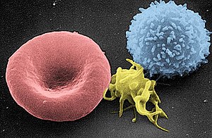
From left to right: erythrocyte, thrombocyte, ...

Image via Wikipedia

Blood can be thought of as a complex chemical solution in which various solids and gases are dissolved, and move simultaneously with the other constituents of blood. As such, the chemical and physical properties of blood are homeostically regulated to fall within specific ranges in the healthy individual. In the disease state, these values are displaced in characteristic ways – allowing the clinician and pathologist to use blood tests to make diagnoses. Blood pH is optimally 7.35 – 7.45 or slightly alkaline. The concentration of dissolved blood gases falls within specific physiological ranges for partial pressures for oxygen, carbon dioxide, as well as bicarbonate and other gases. Oxygen is transported primarily via hemoglobin in red blood cells, while CO2 is transported in solution in the plasma.

[Image shows, left to right, a red blood cell, a platelet, and a small white blood cell, under scanning electron microscopy. – Doc Cindy]

Coming next: blood collection

Copyright © 2011 Peter Farmer

Enhanced by Zemanta
Posted in Contributors, Equipment, Hemocytometer/hemacytometer, Medical archives, Microscope, Perennial Favorites, Pete Farmer, Pneumonia, Sore throat, Strep throat | Tagged , , | Leave a comment

Insulin, thyroxine, and biotech: feasible or fantasy?

Tomorrow’s headline: Hospitals Flooded with Patients Suffering Racing Hearts  – Source Traced to Bioengineered Bacteria. Can Experts Reverse the Epidemic Before More Succumb?

* * *

Today both KF and JW ask about an online article do-it-yourself biotech. Is it actually feasible to duplicate big pharma’s success in producing bioengineered insulin and thyroxine in your own laboratory?

For the technically minded, check out the article they refer to at http://www.indiebiotech.com/?p=135.

While I don’t like to say anything is impossible (are not all things possible with God?), I would estimate the odds of successful production of either product in a home laboratory as close to nil. The article author’s description, while fairly detailed, pales in comparison to what would actually be involved. Perhaps a team of motivated, imaginative, unrestricted, well-funded PhD-level biochemists could concoct these proteins in their basement after a decade of research, but as for counting on this option for treating diabetes and hypothyroidism at TEOTWAWKI, I would not even consider it.

The specific deficiencies the article addresses (lack of insulin in diabetes and lack of thyroxine in hypothyroidism) are really quite separate problems. The thyroid issue is far easier to solve, by resorting to animal sources (and if there aren’t enough animal or human sources around, we’re in big trouble). The thyroid issue is also much simpler because the hormone is effective when taken orally, whereas insulin is not absorbable through the GI tract. Animal thyroid works quite well in humans and thyroid pills should be reasonably stable for years (or perhaps decades) to come. Also, if for some reason thyroid hormone cannot be taken orally, it could be injected (as was done in the late 1800s). Thyroid transplants are theoretically possible but with oral replacement therapy readily available, it has not been a necessity.

The insulin situation is a much greater challenge. It is true that insulin is produced biosynthetically. Recombinant DNA technology now allows for the efficient production of human insulin (which is not immunogenic, like bovine or porcine insulin). However, replicating this technology would be prohibitively difficult. Harvesting insulin from animal pancreatic tissue would be problematic enough.

And what if the recombinant DNA-containg E. coli inoculated the GI tract of researchers? If the engineered bacteria became established as the dominant flora and spread to other family members, the secreted hormones could cause devastating effects. Bacteria producing unquantified amounts of levothyroxine within the human colon could easily cause a condition resembling thyroid storm, a potentially fatal condition.

Although I value imaginative solutions to medical problems, I would classify basement lab production of bioengineered hormones as a pipe dream. (Thanks, KF and JW, for asking.)

On the other hand, if SemBioSys wants to share their secrets of transgenic insulin-producing safflower plants with me (genetic freeware?), I’ll be glad to spread the word.

Please read on for comments from the original article. And how about some input from the other professionals on this site?

Enhanced by Zemanta

Featured image: insuline crystals

Posted in Diabetes Mellitus Type II, Equipment, Hypothyroidism, Insulin, Medical archives, Perennial Favorites, Site Map, Thyroid disease, Thyroid preparations | Tagged , , , , , , , | 9 Comments

Week 21 – Question of the Week: How did your garden grow?

Week 21: 2011-08-26

Labor Day is just around the corner, the end of summer (if not the heat).  At least in Ohio, swimming pools close, amusement parks shut down, and we have only a few more weeks of home-grown corn and fresh tomatoes.

With autumn soon upon us: what have you learned from your garden this year?  What tips can you share with our readers?

Comments, questions, and suggestions are all appreciated.  Submit your response in the block below.

– Doc Cindy

Enhanced by Zemanta
Posted in Question of the Week | Tagged , , , , | 1 Comment

Earthquakes and Ingenuity

At 1:55 p.m. EDT, just before I went out the door, my secretary asked, “Did you feel that?”

At 1:57 I heard Rush Limbaugh’s stand-in say they’d felt an earthquake in New York, with reports soon following of a quake centered near Washington, D.C. Apparently sensitive individuals as far northwest as Akron, Ohio felt it as well.

The Pentagon, the Capitol, the city of D.C. – all evacuated.

Usually these “minor” events take on a bit of a holiday atmosphere. The danger appears minimal, and a little time off work is always welcome. And fortunately, most earthquakes do not cause widespread injury.

However, today’s quake can serve as a wake-up call for those of us less familiar with the aftermath of serious earthquakes.

The CDC offers a starting point for earthquake preparedness at: http://www.bt.cdc.gov/disasters/earthquakes/. The site offers good advice for citizens on what I would call a “controlled disaster,” one in which neither mass panic nor significant injury occurs. Today is a good day to print it out, read it, follow it, and store it. (Will your computer be working when things go bad?)

Readers of Armageddon Medicine may want to investigate further regarding severe injuries that often occur with a serious earthquake.  The CDC’s bioterrorist site on Crush Injuries and Crush Syndrome (see http://www.bt.cdc.gov/masscasualties/blastinjury-crush.asp) estimates the incidence of crush syndrome as 2-15% of quake-related injuries, with half the affected individuals developing acute renal failure, and half of them requiring dialysis. Clearly, this is ICU-level care involving an entire medical team. Professionals may want to think through how to handle such a situation without back-up care. The death rate of patients suffering crush syndrome could easily approach 50%.

After the Haiti quake, of those treated at a hospital, the CDC reports:

The most common injury-related diagnoses were fractures/dislocations, wound infections, and head, face, and brain injuries. The most common surgical procedures were wound debridement/skin grafting, treatment for orthopedic trauma, and surgical amputation. Among patients with earthquake-related injuries, the most common mechanisms recorded were cut/pierce/struck by an object and crush.

(See http://www.cdc.gov/mmwr/preview/mmwrhtml/mm5951a1.htm)

Blast injuries are further enumerated and described at: http://www.bt.cdc.gov/masscasualties/blastinjuryfacts.asp.

Some of our professionals have visited Haiti after the 2010 earthquake. Would any of you care to comment (anonymously, or otherwise) on what you saw or experienced while there?

Or how about this question: what is the most complex medical situation that you have successfully handled without benefit of X-rays, labs, nursing, supplies, or back-up – a real end-of-the-world-as-we-know-it-scenario?

Enhanced by Zemanta
Posted in Bioterrorism, Disaster Relief, Medical archives, Perennial Favorites, Preparation | Tagged , , , , | 3 Comments

Week 20 – Question of the Week: How does 9/11 compare with what’s on the horizon?

Week 20: 2011-08-19

September 11, 2001 my brother was in NYC – not in the midst of the action, but close enough to see the buildings on fire.  He high-tailed it out of the city as quick as he could.  Chaos and confusion were the order of the day.  Thankfully, he made it home safe and sound.

But I wonder: does 9/11 hold a candle to the trouble ahead?

Comments, questions, and suggestions are all appreciated.  Submit your response in the block below.

– Doc Cindy

Enhanced by Zemanta
Posted in Medical archives | Tagged , , , , , | 4 Comments

Photo Quiz Answer – Q.004

Photo Quiz Answer– Q.004 – August 12, 2011  

This was an easy one. Don’t let the patient history throw you off. Though he may have been stung by a bee while camping, this problem is unrelated. 

None - This image is in the public domain and ...

Image via Wikipedia

The image depicts the typical “bull’s-eye” lesion of erythema migrans, the most typical finding of Lyme disease, present in 80% of cases.

Bee stings do not cause this ring-like pattern. Whether the patient needs a bee sting kit is entirely unrelated to this problem. Anyone who experiences shortness of breath, hives, or other symptoms of anaphylaxis needs a bee sting kit. Even if this were a large local reaction to a bee sting, it would not be considered an allergy and would not require a bee sting kit. One exception would be a person prone to repeat stings, possibly a bee-keeper. My own neighbor died of multiple bee stings, though he was not allergic to bees.

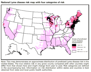
Erythema migrans occurs as a result of infection with the bacterium Borrelia burgdorferi, carried by deer (blacklegged) ticks, pictured above. Patients are frequently unaware of a tick bite and may give no history of exposure. The ticks may be as small as the head of a pin. Other early symptoms include fever, headache, and fatigue. Untreated, the infection may spread to the joints, heart, or nervous system. The infectious is not contagious from human to human.

National Lyme disease risk map with 4 categori...
Image via Wikipedia

Treatment begins with prevention. Anyone living in high risk areas (particularly the eastern United States) should be very cautious about walking through high grass or low bushes in wooded areas, particularly in the summer when the deer ticks are most active. Though they normally feed on deer, birds, and mice – the common inhabitants of such a region – they can also feed on humans, horses, cats, and dogs. A tick must actually attach and feed to transmit Lyme disease. Therefore, wearing long pants and long sleeves when in the woods is essential, as is removing any tick as soon as it is noticed, preferably before feeding has occurred. Use insect repellant (containing DEET) and check yourself frequently.


Anyone who demonstrates a typical bulls-eye rash and has had any likelihood of a tick bite should be treated with antibiotics. (Doxycycline for adults and children age 9 and above; amoxicillin or cefuroxime for younger children and pregnant or nursing women. Treat for 10-21 days.)   

Prepping summary:

1. Procure long pants and long-sleeved clothing for all family members.

2. Include insect repellant containing 10-30% DEET

3. Learn to recognize the deer tick

5. Include doxycycline and amoxicillin in your prepping supplies.

6. Read more about Lyme disease at: http://www.mayoclinic.com/health/lyme-disease/DS00116

Enhanced by Zemanta
Posted in Bites-insect-bug-tick, Fatigue, Fever, Injuries, Lyme disease, Medical archives, Perennial Favorites, Photo Quiz Answers, Rash, Skin infection, Tick-borne illness | Tagged , , , , , | 3 Comments